
Note that the Obturator Externus is not illustrated, but would be deep to pectineus and superior to adductor musclesĬreative commons source by Beth ohara, edited by Thomas Burnell They are innervated by the obturator nerve.ĭiagram - Medial compartment of the thigh. These muscles are generally involved in hip adduction. In addition to extension of the hip and flexion of the knee, it can medially rotate the thigh. When looking at the back of the thigh, it can be difficult to differentiate between semintendinosus and semimembranosus - semimembranosus is the most medial of the hamstrings (both 'm' words). It attaches to the medial tibial condyle. Like the the other hamstrings, it originates from the ischial tuberosity. This is the most medial of the posterior muscles.

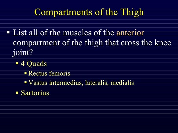
When looking at the back of the thigh, it can be difficult to differentiate between semintendinosus and semimembranosus - semitendinosus is the one with a very long tendon at the bottom that attaches to the medial tibia. It is innervated by the tibial branch of the sciatic nerve. It is primarily responsible for flexion of the knee joint and extension of the hip, but can also medially rotate the thigh. It originates from the ischial tuberosity and attaches to the medial tibia. This muscle lies medial to the biceps femoris. It can contribute to some lateral rotation of the hip and the knee, as well as extension of the hip and flexion of the knee. The two heads are innervated by two different branches of the sciatic nerve: the long head by the tibial branch, and the short head by the common fibular branch. This tendon can be palpated on the lateral aspect of the posterior knee. Together, they insert into the head of the fibula. The long head originates at the ischial tuberosity, whilst the short head originates at the linea aspera of the femur. They are innervated by the sciatic nerve.ĭiagram - Posterior compartment (Hamstring) muscles on the right leg highlighted in redĬreative commons source by OpenStax College, edited by Thomas Burnell Collectively, these muscles are known as the hamstrings and are involved with extension of the hip and flexion of the knee. Thankfully, the posterior compartment is simpler. This is an Advert - we use these to keep SimpleMed free! If you see something you like, please click on it - it supports the site :) They contribute to flexion of the hip joint. This muscle is a combination of both the psoas major and iliacus muscles, hence the name. It originates from the ilium and attaches to the medial tibia. This muscle makes up the fourth muscle of quadriceps femoris. Vastus Medialis – originating from the medial lip of the linea aspera.Vastus Intermedialis – originating from the anterior surface of the femoral shaft.Vastus Lateralis – originating from the lateral lip of the linea aspera.They all attach to the patella via the patella tendon. These muscles are responsible for extension of the knee joint. There are three vastus muscles that contribute the quadriceps femoris group of muscles. It’s placement within the body allows it to flex and adduct the hip joint. This is a flat muscle that runs from the anterior surface of the pubis to the pectineal line of the femur. It flexes, abducts and laterally rotates the hip joint and flexes the knee joint. It runs from the anterior superior iliac spine and runs inferomedially to the medial tibia. It is long, thin and is the most superficial of the anterior compartment muscles. This muscle is the longest in the body and runs in an oblique fashion across the anterior thigh. Note that the vastus intermedialis tucked underneath the rectus femoris and is, therefore, not visibleĬreative commons source by OpenStax College Generally speaking, these muscles act to flex the leg at the hip joint and can also act on the knee joint.ĭiagram - Muscles of the anterior thigh.

The muscles of the anterior compartment of the thigh are innervated by the femoral nerve. Pink represents the anterior compartment, green is the medial compartment and blue represents the posterior compartment For more information, please check out our Anatomical Terminology Article.ĭiagram - The fascial compartments of the thigh. It is also very important when studying muscles that you are comfortable with anatomical terminology, as a lot is used in these articles. These compartments are separated by layers of fascia.

There are many muscles in the thigh, however, they can be categorised into the anterior, posterior and medial compartments. The muscles of the thigh are responsible for a variety of movements, acting on both the hip and the knee joint.


 0 kommentar(er)
